PET/CT imaging of pancreatic carcinoma targeting the “cancer integrin” αvβ6
Neil Gerard Quigley, Norbert Czech, Wolfgang Sendt & Johannes Notni
αvβ6-Integrin is exclusively expressed by epithelial cells and plays an important role for invasion and metastasis of carcinomas. We found a high expression of β6 on tumor cells in 88% of nearly 400 specimens of pancreatic ductal adenocarcinoma (PDAC) primaries and in virtually all metastases [1]. We earlier reported a series of 68 Ga- and 177Lu-labeled αvβ6-integrin-specific cyclic nonapeptides, but found that despite some showed a good tumor-to-background contrast in rodent models, tumor accumulation was ultimately too low for a successful clinical transfer [2]. We hypothesized that trimerization might result in elevated target-specific uptake and prolonged retention and thus elaborated a trimerized αvβ6-specific 68 Ga-peptide named 68 Ga-Trivehexin.
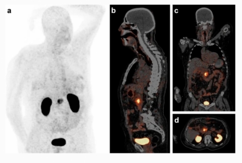
The image shows a 68 Ga-Trivehexin PET/CT of a male patient (82 y, 89 kg) with a histologically confirmed PDAC in the pancreatic head (87 MBq, 70 min p.i., acquisition time 25 min, 0.7 mm/s, 3 min/bed position; anterior MIP (a) scaled to SUV 12; PET in slices (b–d) to SUV 10). Apart from the PDAC lesion (SUVmax = 13.1), prominent signals are observed only in kidneys and urinary bladder due to renal excretion. No relevant uptake is seen in lungs, stomach, liver, and intestines. In light of a limited value of [18F]FGD-PET for early detection of PDAC [3], we anticipate that 68 Ga-Trivehexin will have a clinical value in this setting, besides the potential applications for fibrosis and other carcinomas (head-and-neck squamous cell, lung adenocarcinoma, colon, cervical, mammary) which have been addressed previously by αvβ6-integrin targeted PET-radiopharmaceuticals [4,5,6].


